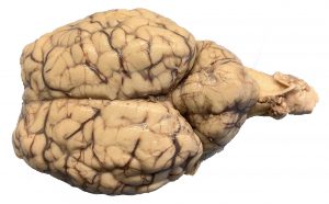
Image of Sheep Brain. Northern Virginia Community College by H.W. is licensed under CC BY 4.0
Case Study: “Stumbling Stan”
It’s a busy night in the city’s emergency department (ER). Paramedics bring in a 20-year-old man named Stan by ambulance. He is writhing in pain due to a terrible headache, can’t answer questions properly (he doesn’t know today’s date), doesn’t understand where he is, and is experiencing sudden loss of vision. As the paramedics move him into a hospital bed, he reels and vomits. He is conscious but confused. His speech is slurred. The physician orders an MRI of his head to evaluate the brain and surrounding structures, and a cranial nerve exam.
Objectives
By the end of this lab, students will be able to….
The Brain:
- Identify and describe the function(s) of parts of the brain on both an image and model.
- Identify the starred (*) structures from the terminology checklist on a sheep’s brain.
- Identify the meninges and the spaces created by the meninges on both an image of the brain and on a brain model.
- Trace the flow of cerebrospinal fluid (CSF) through the brain from its origin in the choroid plexus to where it collects in the superior sagittal sinus.
- Given the location of a brain lesion of a specific part of the brain, predict the effect the lesion will have on brain function.
The Cranial Nerves
- Identify and describe the basic function(s) of the 12 cranial nerves on an image or model.
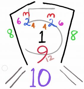
Diagram of the 12 cranial nerves. Northern Virginia Community College by Gillian Backus is licensed under CC BY 4.0
- Categorize each cranial nerve as sensory, motor, or both (mixed).
- Evaluate cranial nerve function from the results of common cranial nerve tests.
Materials
Brain model
- CSF sagittal section model
- terminology labels and sticky tack
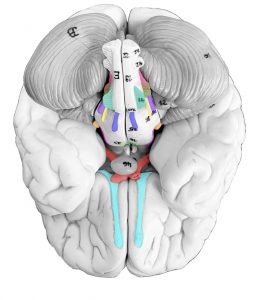
Inferior view of brain with colored cranial nerves. Northern Virginia Community College by Heidi Wangerin is licensed under CC BY 4.0
- ventricle model
- disposable gloves
- dissecting tray and foam pad
- dissection tools (blunt probe, scalpel, fine probe, scissors)
- lab apron
- safety goggles
- sheep brain
- slotted spoon
- colored pencils
- 3-4 cotton balls
- activator
- penlight
- small, labelled vials filled with 3 different scents
- Snellen eye chart
- tuning fork
Try the lab!
Instructor Resources
Need something? Have comments? Fill in the contact form below:
![]()
A&P 1 OER Lab Manual © 2022 by H. Wangerin, P. Rodgers, G. Backus is licensed under CC BY-NC-SA 4.0 ![]()
![]()