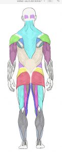
Colored Muscles of the Back © 2022 by H. Wangerin is licensed under CC BY-NC-SA 4.0
Case Study: “Too Tired to Stand”
You are working as a nurse’s assistant in a pediatrician’s office. As you bring the next patient, a 5-year-old boy named Jose, back to the exam room, he trips, and you catch him. His parents comment that he falls frequently. You observe how he moves and notice that he walks on his toes and uses his arms for balance. You notice that his calves appear larger than the average child of his age. When you get to the exam room, he is mildly out of breath. After questioning the parents, you learn that Jose has trouble in school and suspect he has a learning disability. The parents are concerned that Jose has trouble running and jumping. He also frequently complains of muscle pain. When you question Jose about the pain, he comments that today his shoulder is sore. You ask him to show you how he ties his shoes. After he is done, he places his hands on his ankles and “walks” them up to his shin, knee and then thigh to stand upright. (This method of standing up is called “Gower’s sign”.) The pediatrician will want to see that you include all the muscles that might be affected in your notes. It might be wise to review the major muscles of the body!
Background
There are over 600 muscles of the human body. Learning their names, locations and actions can be intimidating. In this lab we will focus on a select subset of muscles that contribute to major body movements. Skeletal muscle usually attaches to two different bones, one called the origin and the other at the insertion, and typically spans a joint. Muscles connect to the bone by a cord of dense connective tissue, called a tendon. When the muscle contracts, it gets shorter as it pulls one bone (insertion) toward the other bone (origin). Thus, in most cases the origin is stationary while the insertion moves toward the origin.
Objectives
By the end of this lab, students should be able to…
- Identify the given muscles of the body on a model or image.
- Identify a muscle on the body in anatomical position as being anterior, posterior, medial, lateral, superficial and/or deep.
- Identify the major actions of each muscle listed using terms like flexor, extensor, adductor, abductor, rotator etc.
- Identify the origin and insertion points of selected muscles (with an asterisk *) using terms from your axial and appendicular lab.
- Define, identify, and label a tendon, ligament, and aponeurosis.
- Given the origin and insertion of selected muscles (with an asterisk *), predict the action of a muscle.
Terminology
*NOTE* Below is a list of the muscles we ask our students to learn. Those with an asterisk are those which students must learn origin, insertion, and action as well.
Muscles of Facial Expression
- epicranius (occipitofrontalis)
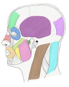
Colored image of lateral view of selected muscles of the head © 2022 by H. Wangerin is licensed under CC BY-NC-SA 4.0
- frontalis
- occipitalis
- nasalis
- orbicularis oculi
- orbicularis oris
- platysma
- zygomaticus
Muscles of Chewing
- buccinator
- lateral pterygoid
- masseter*
- temporalis
Muscles of the Head, Neck, and Vertebral Column
- erector spinae
- spinalis (medial)
- longissiumus (intermediate)
- iliocostalis
- sternocleidomastoid *
Muscles of Respiration
- diaphragm*
- external intercostals
- internal intercostals
Muscles of the Abdominal Wall
- external oblique
- internal oblique
- rectus abdominis*
- transverse abdominis
Muscles of the Pectoral Girdle and Arm (Humerus)
- deltoid*
- latissimus dorsi*
- pectoralis major*
- rhomboid rotator cuff muscles infraspinatus* subscapularis* supraspinatus*
- serratus anterior
- teres major
- trapezius (superior section)*
Muscles of the Forearm (Radius & Ulna)
- biceps brachii*
- brachialis*
- brachioradialis
- pronator teres
- supinator
- triceps brachii*
Muscles of the Carpus and Hand
- extensor carpi radialis longus
- extensor digitorum
- flexor carpi radialis
- flexor carpi ulnaris
- flexor digitorum superficialis
Muscles of the Hip and Thigh (Femur)
- adductor muscles
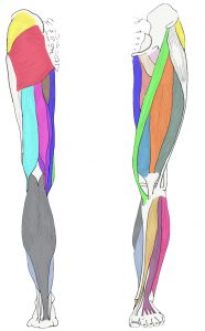
Colored image of posterior and anterior surface of the muscled leg © 2022 by H. Wangerin is licensed under CC BY-NC-SA 4.0
- adductor longus
- adductor magnus
- gluteus maximus
- gluteus medius*
- gracilis
- iliopsoas
- iliacus
- psoas major
- sartorius
Muscles of the Tibia and Fibula
- hamstring group
- biceps femoris*
- semitendinosus
- semimembranosus
- quadriceps femoris
- rectus femoris*
- vastus lateralis
- vastus medialis
- vastus intermedius
Muscles of the Ankle and Foot
- extensor digitorum longus
- fibularis longus (peroneus longus)
- gastrocnemius*
- calcaneal tendon (Achilles’ tendon)
- soleus
- tibialis anterior*
Materials
- Muscle models of entire body
- Muscle models of head and neck and trunk
- Muscle models of human arm and human leg
Let’s do the lab!
Instructor Materials
Is Something Missing? Want to send us comments? Fill in the contact form!
A&P 1 OER Lab Manual © 2022 by H. Wangerin, P. Rodgers, G. Backus is licensed under CC BY-NC-SA 4.0 ![]()
![]()