Case Study: “Hips of Steel”
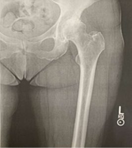
Radiograph of hip prior to surgery © 2022 by Heidi Wangerin is licensed under CC BY-NC-SA 4.0
Summer is peak activity in the physical therapy office where you volunteer as an assistant to the physical therapist. The next patient is a 70-year-old female who has just had a second hip replacement surgery. She is 30 days post-op and is coming in for an evaluation of her hip and for an exercise and strengthening plan during her rehabilitation. Before she comes in you compare her before and after radiographs and realize that the anatomy of her hip has changed. The physical therapist starts quizzing you on the bones of the lower limb. You accidentally identify the femur as the humerus. The physical therapist gently suggests you review the appendicular skeleton to identify the main bones of the upper and lower limb on the articulated skeleton.
Objectives
By the end of the lab, students should be able to…
- Identify the bones and markings on the terminology checklist for the appendicular skeleton.
- Correctly determine whether each of the following disarticulated bone is right or left: scapula, humerus, femur, and tibia.
Materials
- articulated skeletons
- disarticulated skeletons
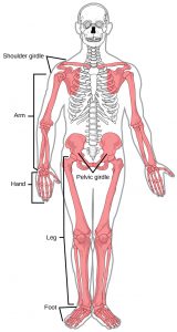
Highlighted bones of the appendicular skeletons © 2022 by Heidi Wangerin is licensed under CC BY-NC-SA 4.0
- ability to label skeletons (we suggest making laminated labels of the terminology and using sticky tack)
Terminology for this lab
*Note* We recognize that each Anatomy and Physiology lab may have a different list of terms. This is the list of terms we have decided to use for our 100-level undergraduate anatomy and physiology course for allied health students.
Bones and Bone Markings of the Upper Limb
- clavicle (acromial (lateral) end, sternal (medial) end)
- scapula (acromion, coracoid process, glenoid cavity, infraspinous fossa, spine, subscapular fossa, supraspinous fossa)
- humerus (capitulum-lateral condyle, coronoid fossa, deltoid tuberosity, greater tubercle, head, lesser tubercle, neck, olecranon fossa, trochlea-medial condyle)
- radius (head, radial styloid process, radial tuberosity, ulnar notch)
- ulna (coronoid process, head, olecranon process, radial notch, trochlear notch, ulnar styloid process)
- carpals (trapezium)
- metacarpals
- phalanges (name and number of each)
Bones and bone markings of the lower limb:
- femur (fovea capitus, greater trochanter, head, lateral condyle, lateral epicondyles, lesser trochanter, medial condyle, medial epicondyles, neck, patellar surface)
- patella
- tibia (lateral condyle, medial condyle, medial malleolus, tibial tuberosity)
- fibula (head, lateral malleolus)
- tarsals (calcaneus, talus)
- metatarsals
- phalanges (name and number of each)
- pelvis (Os Coxae or coxal) which is made of …..
-
- ilium (acetabulum, anterior inferior iliac spine, greater sciatic notch, iliac crest)
- ischium (ischial spine, ischial tuberosity)
- pubic bone (obturator foramen, pubic arch, pubic symphysis)
Do the lab!
Appendicular Skeleton Lab (Su23)
Instructor Resources
Have comments or questions? Need Something? Fill in the Contact Form below:
![]()
A&P 1 OER Lab Manual © 2022 by H. Wangerin, P. Rodgers, G. Backus is licensed under CC BY-NC-SA 4.0 ![]()
![]()