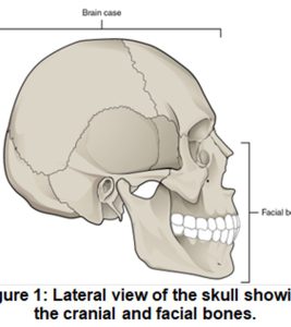
Image of Lateral View of Skull and Jaw. OPEN STAX
Case Study: “Car Accident Woes”
You are wondering what path to take in the health sciences. Becoming a dental assistant or dental hygienist interests you. Your advisor suggests that you shadow someone in these fields to see if you would like to pursue either as a profession. A family friend, Dr. Hernandez, has invited you to come to her office for a day to shadow her and the dental hygienist. The patient sustained a fractured jaw following a car accident when the air bag in the passenger seat where he was sitting failed to deploy. Thankfully, he was wearing a seat belt, which probably saved his life. His neck, chest, and face are all sore. His jaw appears disfigured. You are studying bones in your A&P class and pull out your notes to review which bones might be involved. You think that the jaw might be correctly termed the mandible, but the maxillae could also be involved. Dr. Hernandez confirms that the mandible is involved in the fracture, but the zygomatic and temporal bones are as well. She describes the fractured processes of the bones and discusses possible nerve damage. She asks you what a general term is for a bony passageway for nerve and blood vessels. You quickly answer, “a meatus.” She replies, “you are very close! But a better term would be ‘foramen’”. Slightly embarrassed, you decided it might be a good time to review general bone marking terms!
Objectives:
By the end of this lab, students will be able to:
- Define and apply the bone marking terminology to describe bones of the skeleton.
- Identify the bones and markings on the terminology checklist for the axial skeleton.
- Apply what you have learned: evaluate why the features of the fetal skull are different from an adult skull.
- Apply what you have learned: Given bone marking descriptions, properly identify a bone.
*Note* This lab focuses on the Axial skeleton, namely the bones of the skull vertebral column, ribs and sternum. Students also learn the names of the fontanels in a fetal skull. A second lab focuses on the bones and bone markings of the appendicular skeleton.
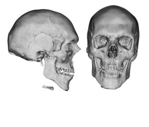
View of skull © 2022 by Heidi Wangerin is licensed under CC BY-NC-SA 4.0
We recognize that not every school has students learn the same bones and bone markings. To that end, we have included here our terminology list. As an OER resource, you are welcome to adapt this resource to suit your students’ and your school’s needs.
Bone and Bone Markings of the Skull
Bone markings are noted in parentheses.
- frontal bone (supraorbital foramen (notch) )
- parietal bone
- temporal bone (external acoustic meatus, mandibular fossa, mastoid process, styloid process, zygomatic process)
- occipital bone (foramen magnum, occipital condyle)
- ethmoid bone (cribriform plate, crista galli)
- sphenoid bone (greater wings, lesser wings, optic canal, sella turcica)
- mandible bone (coronoid process, mandibular condyle, mandibular foramina, mandibular notch, mandibular ramus, mental foramen)
- maxillae bone
- palatine bone
- zygomatic bone (temporal process)
- lacrimal bone
- vomer bone
- nasal bone
- inferior nasal concha
- sutures (coronal suture, lambdoid suture, sagittal suture, squamous suture)
- fetal skull (anterior fontanel, posterior fontanel)
General Features of a Vertebra
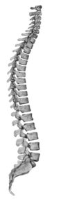
Lateral View of Vertebral Column © 2022 by Heidi Wangerin is licensed under CC BY-NC-SA 4.0
- body
- inferior articular facet
- inferior articular process
- intervertebral foramen
- lamina
- pedicle
- spinous process
- superior articular facet
- superior articular process
- transverse process
- vertebral arch
- vertebral foramen
Regions and Specific Bone Markings of the Vertebral Column
Students should be able to identify all the general structures of a vertebral bone referenced in the list above plus specific terms as noted below, for each type of vertebra)
- Cervical C1-C-7 (transverse foramen, C1 = atlas, anterior arch, C2= axis, dens)
- Thoracic T1-T12 (inferior costal facet, superior costal facet)
- Lumbar L1-L5
- Sacrum (sacral canal, sacral foramen, sacral hiatus)
- Coccyx
Thoracic Cage
- sternum (body, manubrium, xiphoid process)
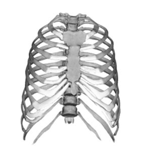
Frontal View of Rib Cage © 2022 by Heidi Wangerin is licensed under CC BY-NC-SA 4.0
- rib (head, costal cartilage, neck, shaft, tubercle)
- false ribs (8-12)
- floating ribs (11-12)
Bones of the Anterior Neck
- hyoid bone
Materials needed for this lab
- articulated skeletons
- disarticulated skeleton (particularly skull, vertebrae from cervical, thoracic, lumbar and sacral regions, and ribs and sternum
- fetal skull models
Let’s do the lab!
Instructor Resources
Want to know more? Have a comment? Please write to us!
![]()
A&P 1 OER Lab Manual © 2022 by H. Wangerin, P. Rodgers, G. Backus is licensed under CC BY-NC-SA 4.0 ![]()
![]()