
Case Study: “The Case of the Wobbly Gymnast”
Sprained Ankle © 2022 by Heidi Wangerin is licensed under CC BY-NC-SA 4.0
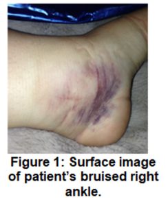
You are a pediatrician for a sports medicine practice. Your next patient is a 12-year-old gymnast. When she dismounted the balance beam during practice, she landed and inverted her right ankle. She has bruising around the lateral aspect of her right ankle, swelling and acute pain around her lateral malleolus, and limited range of motion. She is concerned that she has fractured her ankle and asks you about some common ankle injuries.
You tell her that these symptoms may suggest a sprain, a strain, or a fracture. Sprains and strains involve the stretching of the ligaments, tendons and muscles supporting a joint. A fracture involves the breaking of a bone (usually) at the joint. A less common injury is a high ankle sprain, also referred to as syndesmotic injury. A syndesmosis is a fibrous joint that unites the tibia and fibula by a sheet of connective tissue called the interosseous membrane. In a high ankle sprain, the interosseous membrane is overstretched which causes pain and instability between the tibia and fibula. You explain that ankle injuries are quite common and can range from mild to severe. Moreover, inversion injuries (rolling the ankle outward) are much more common than eversion injuries (rolling the ankle inward) because the medial side of the ankle is more stable due to the large deltoid ligament and the presence of the bony medial malleolus of the tibia. The lateral side of the ankle consists of the much thinner and more delicate fibula.
Background
You have 206 bones in your body. Every single one, except for the hyoid bone in the neck region, is connected to another bone. Joints, or articulations, are the meeting points between bones. Joints vary in the structure and degree of movement. Joints are classified as synarthrotic (non-mobile and highly stable), amphiarthrotic (slightly moveable) or diarthrotic (freely mobile). Here, we focus on the diarthrotic joints, the ones that permit movement. You will learn about the variety of types of movement possible at different types of diarthrotic joints — from the neck and head, to the shoulder and hip, to the elbow and knee, to the wrists and ankles, to the fingers and toes. Let’s get moving!
Note: The 2023 version of this lab focuses on synovial joints. References to synarthrotic joints and amphiarthrotic joints have been removed, in the interest of time.
Objectives
By the end of the lab, students will be able to….
- Define the terms on the terminology checklist and apply these terms to specific joints, including the knee joint.
- Classify a given joint based on its anatomical structure as fibrous, cartilaginous, or synovial.
- Classify a given joint based on its function as a diarthrosis, amphiarthrosis or synarthrosis.
- Define and demonstrate the different types of movements possible at synovial joints.
- Name the structural components of a typical synovial joint.
- Given an image (or description) of a joint position, determine the joint movement (or description), articulating bones, (and muscle action if muscles have been covered).
This lab occurs in Stations, using a jigsaw format.
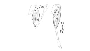
Flexion of a elbow joint and contraction of biceps and relaxation of tricepts by Heidi Wangerin is licensed under CC BY 4.0
Materials
- Station 1: skull, atlas bone, axis bone, head muscle model, spine bone model, small body muscle model
- Station 2: scapula bone, humerus, shoulder joint model, small body muscle model, arm muscle model
- Station 3: humerus, ulna, radius, arm muscle model
- Station 4: femur, pelvic model, leg muscle model, hip joint model
- Station 5: tibia, fibula, patella, femur, leg muscle model, knee joint model
- Station 6: articulated foot, fibula, tibia, leg muscle model, articulated hand, and arm muscle model
- completed Muscles Chart from the Muscles lab
- completed bones study guides, with bone markings labeled
- body in motion images
- joints worksheet blank vs 3
Try the lab!
Synovial Articulations Lab Su23)
Note: The 2023 version of this lab focuses on synovial joints. References to fibrous joints and amphiarthrotic joints have been removed, in the interest of time.
Instructor Resources
- Memo Articulations 2023
- Instructor PPT Articulations (2023, as pdf)
Got questions or comments? Need something? Fill in the contact form below:
![]()
A&P 1 OER Lab Manual © 2022 by H. Wangerin, P. Rodgers, G. Backus is licensed under CC BY-NC-SA 4.0 ![]()
![]()