Case Study: “The Mysterious Femur Fracture”
An 86-year old woman named Therese has been referred to you following a fracture at the neck of her left femur (at the greater trochanter, near the proximal epiphysis). She fractured her femur after slipping in the shower (Figure 1).
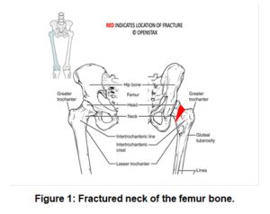
Femur Fracture at proximal end. OPENSTAX
Typically, femurs will only fracture under a tremendous amount of stress. It is unusual that she fractured this bone after a fall from standing height.
Objectives
By the end of this lab, students will be able to…
- Identify the parts of a long bone.
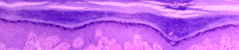
- Differentiate among the types of cartilage based on their morphology, location, and/or function.
- Compare and contrast the structure, function, and location of compact and spongy bone.
- Identify the structures of an osteon from an image, model or microscope slide.
- Trace the flow of nutrients through compact bone using appropriate terminology from an external blood vessel to an individual lacuna (and osteocyte).
- Explain the organic and inorganic components of the extracellular matrix of compact bone and briefly describe the function of each.
Materials
- Images in the Instruction Guide
- Compound microscope (one per student)
- Prepared slides of hyaline cartilage, fibrocartilage, and elastic cartilage or use Histology Guide
- Model of an osteon and/or laminated image of a model of an osteon.
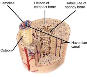
Image of Compact and Spongy Bone, OPENSTAX
- Acid-soaked chicken bone
- Baked chicken bone
- Fresh chicken bone
Want to learn more? Try the Lab!
Instructor Resources
Want to know more? Have comments? Please fill in the contact form below!
![]()
A&P 1 OER Lab Manual © 2022 by H. Wangerin, P. Rodgers, G. Backus is licensed under CC BY-NC-SA 4.0 ![]()
![]()