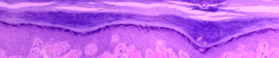
Pseudostratified Columnar Epithelium © 2021 by H. Wangerin is licensed under CC BY-NC-SA 4.0
Case Study: “Case of the Persistent Cough”
You are a physician assistant for a pulmonologist. Your first patient of the afternoon is a 26-year-old male who has been experiencing a painful cough for the past 6 weeks (about 1 and a half months). The patient had been a chain smoker for 4 years and switched to vaping 6 months ago after hearing from friends that it was a “safer” alternative to smoking. An initial pulmonary CT scan showed that his lungs appeared clear. No cancerous masses or fluid accumulation were detected. To further evaluate the patient, you have been asked to assist with a bronchoscopy in which a tiny camera is inserted through the mouth and down into the trachea and lungs. The bronchoscopy will allow you to visually analyze the state of the tissue lining the respiratory tract to obtain a biopsy for histological analysis.
- NOTE* We tend to do this lab over the course of 2 weeks, since there are a lot of slides to learn. We have 6 examples of epithelial tissue and 11 examples of connective tissue! In part I, students study the epithelial tissue types, and start on the connective tissue, such that students learn about 8 different tissue types each week. This lab allows students to investigate the three types of muscle tissue and nerve tissue by looking at images only. They will revisit these two tissue types when they study the muscular and nervous systems respectively.
Objectives
By the end of this lab, students should be able to….
- Define histology.
- List and define the 4 types of tissue in the human body.
- Locate and identify from an image or histology slide the diverse types of epithelial tissue.
- Describe the function and common locations of the diverse types of epithelial tissue.
- If applicable, describe the structural modifications (cilia, microvilli, or keratinization) that contribute to the specific function of each epithelial tissue sub-type.
- Locate and identify from an image or histology slide the several types of connective tissue (11 types).
- Describe the function and common locations of the diverse types of connective tissue.
- On an image or histology slide of connective tissue, identify the ground substance and the diverse types of protein fibers that create the extracellular matrix of connective tissue.
- Describe how protein fibers and ground substance contribute to the function of the different connective tissue types.
- On a picture or histology slide of connective tissue, identify the most common type(s) of cells present.
- Differentiate between the three types of cartilage.
- Locate and identify from an image or histology slide the 3 distinct types of muscle tissue.
- Describe the function and common locations of the 3 diverse types of muscle tissue.
- On an image or histology slide, identify nervous tissue.
- Define an organ.
- Explain the several types of tissue in an organ and how they contribute to the organ’s function.
*By the end of this lab you should have an idea of the typical pattern each tissue has. Do not try to memorize a set image of the tissue types but try to understand the function of a tissue and how it would relate to its morphological features.
Want to learn more? Try the lab!
Materials
- Compound microscope (one per student)
- Dropper bottle of isopropyl alcohol and lens paper for cleaning microscope lenses
- PART I: Prepared slides of simple squamous epithelium, simple cuboidal epithelium, simple columnar epithelium, ciliated pseudostratified epithelium, nonkeratinized stratified squamous epithelium, and transitional epithelium
- PART I or II : Prepared slides of connective tissue to include areolar tissue, adipose tissue, dense regular connective, dense irregular connective, and dense regular elastic tissue.
- PART II: Prepared slides of elastic cartilage, hyaline cartilage, fibrocartilage, compact bone, and blood.
- Prepared slides of smooth muscle, skeletal muscle, and cardiac muscle (not required)
- Prepared slide of nervous tissue (not required)
- Online resource: Excellent histology slides available from Histology Guide website.
Instructor Resources
- Memo Histology
- PPT Histology Part 1 2023
- PPT Histology Part 2 2023 (Supportive Connective Tissue, Muscle and Nervous Tissue)
Not finding what you need? Want to leave a comment? Fill out the contact form below!
![]()
Bio141 OER Lab Manual © 2022 by H. Wangerin, P. Rodgers, G. Backus is licensed under CC BY-NC-SA 4.0 ![]()
![]()