Case Study: “Cancer is Only Skin Deep”
Thick Skin by Heidi Wangerin is licensed under CC BY 4.0
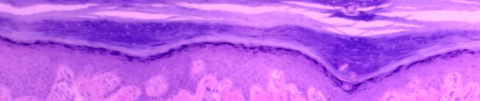
The skin is the largest organ in the body; it accounts for about 15% of your body weight, and contains over 300 million skin cells and 11 miles of blood vessels! Your skin continuously replenishes itself –the outer layer sloughs off and is replaced by new skin cells underneath. You might be surprised to learn that the sloughed off cells account for a substantial percentage of the dust around your house! The many functions of the integument include sensation, protection, and regulation. Many different structures in the skin help to carry out these various functions, including sensory receptors, muscles and glands. The skin itself is made up of the epidermis, the outer layer of cells which is about 0.1 mm thick, and the dermis, the lower layer which is about 4 mm thick. Below these two layers of skin is a deeper layer of connective and adipose tissue called the hypodermis. This layer serves to anchor and connect the skin to the underlying organs.
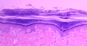
Histology of Carincoma © 2021 by J. Bragg is licensed under CC BY-NC-SA 4.0
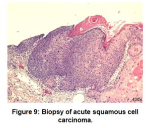
As a nurse working in a dermatology office, patients typically come into the office with various nodules, lesions, and rashes that need to be imaged and/or biopsied to test if they are cancerous. This afternoon a middle-aged female walks into the office with a flat, scaly sore on her leg. The patient has several risk factors for skin cancer including using tanning beds in her teenage years and having fair skin and light-colored eyes. You meet with the patient and make several visual observations of the lesion as well as record her medical history. You mark the location of the lesion on the leg (Figure 1) and take a close-up image of the features and size of the lesion (Figure 2). Working closely with the dermatologist, you biopsy a small piece of the lesion to be sent to the lab and tested for the presence of cancer cells.
Objectives
By the end of this lab, students should be able to….
- Identify the specific tissue type and explain the function of each layer of the epidermis, dermis, and hypodermis.
- Identify, label, and define the function of the structures of the integument from a model or image.
- Identify the layers of the epidermis and dermis from a microscope slide.
- Explain the function and location of the different structures and cells of the epidermis, dermis, and hypodermis.
- Differentiate between thick and thin skin based on appearance, structure, and function.
Materials
- Microscopes (1 per student)
- Prepared slides of thick skin (sole of foot)
- Prepared slides of thick skin (scalp, or skin with hair follicles)
- Skin model
- Terminology labels (with sticky tack)
- Dropper bottle of isopropyl alcohol and lens paper for cleaning microscopes.
Let’s do the lab!
Instructor Resources
How much have you learned?
Once you have finished the lab, try these sample questions. More are found in the link of the pdf above.
- Compare and contrast the features of thick and thin skin.
- Given the description below, predict whether you expect the area to be covered by thick or thin skin.
- An area of the body commonly subjected to high levels of abrasion ______
- Region of the body that releases sweat into a hair follicle _______
- Given the function below, identify the structure (or layer) of the skin which fits this function.
- Sensory receptors that respond to deep touch/pressure. _____________
- Sensory receptors which respond to light touch.______________
- Skin layer which houses capillaries to allow nutrients to diffuse efficiently into the avascular epidermis. _______________
- Secretes lipids which serve as a waxy barrier to keep the skin moist. _______________
- Secretes a protein rich liquid which produces an odor that is influenced by sex hormones. _______________
- Secretes a liquid that is mostly water and serves to cool the body. ______________
- The histological sample of your patient shows that there is an over-abundance of keratinocytes. Explain the normal function of keratinocytes in the epidermis.
Interested in learning more? Have comments? Need an answer key (Instructors only)? Fill out the form below!
![]()