Case Study: “Assisting the Surgeon”

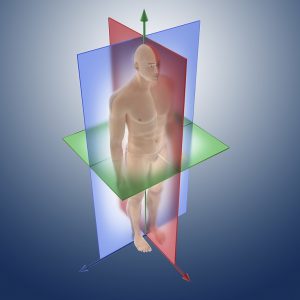
Anatomical orientation planes. OPENSTAX
You are a physician’s assistant working a rotation in the ER. One evening a male patient is rushed into the ER with multiple stab wounds. He is unresponsive and having difficulty breathing. The paramedics provide you with his vitals: heart rate 150 beats/minute (normal = 50-75 beats/minute), blood pressure 80/60 (normal is 120/80), blood oxygen saturation of 90% (normal is 99%). He is immediately placed on oxygen and his injuries imaged to evaluate their extent for the surgical team. Your job is to assess the injuries, describe their location and relative position on the body. It is critical that you record this information accurately for the attending surgeon to operate properly. To do this, we have divided the lab into six activities, designed to introduce you to terms that you need to add to your list of working vocabulary. You will use these terms in activity 7 to fill out a report for the surgeon about this patient. At the end of this lab you will be expected to label models or images without a word bank. You will also need to be able to describe the location of various organs and surface anatomy using appropriate directional terms, again without a word bank. Above is a full body picture showing the location of the three injuries sustained by the patient (Figure 1).
Objectives
By the end of this lab students will be able to…
- Describe the function(s) of each of the 11 organ systems.
- Identify the major organs of each organ system and explain their function(s).
- Display and describe standard anatomical position.
- Name the regions of the body using proper surface anatomy terms.
- Define each directional term and use them to describe the relationship between two structures on a body.
- Describe the three different body planes using directional terms to divide the body into sections.
- Given a body image, determine the body plane that is represented.
- Name and identify the location of the cavities in the body and list the major organs in each cavity.
- Identify and name the serous membranes for the pleural, pericardial, and abdominal cavity.
- Identify the 4 quadrants and the 9 regions of the abdomen. Identify the main organs in each region.
- Apply your knowledge to a medical scenario by describing the surface location, serous membranes penetrated, relative position, plane of section, and abdominal quadrant or region (if applicable) of an injury on a patient.
Interested in learning more? Try the lab!
Materials
- Anatomical Terminology labels
- Torso Model
- Muscle Model
- Labeled Model or image of the human body
- Blunt Probe
- Laminated images (see instructor resources)
- Modeling Clay
- Plastic knife
- Resealable bag with 15 mL of water (dyed blue)
- Laminated images of torso models (see instructor resources)
- Dry Erase marker and eraser
